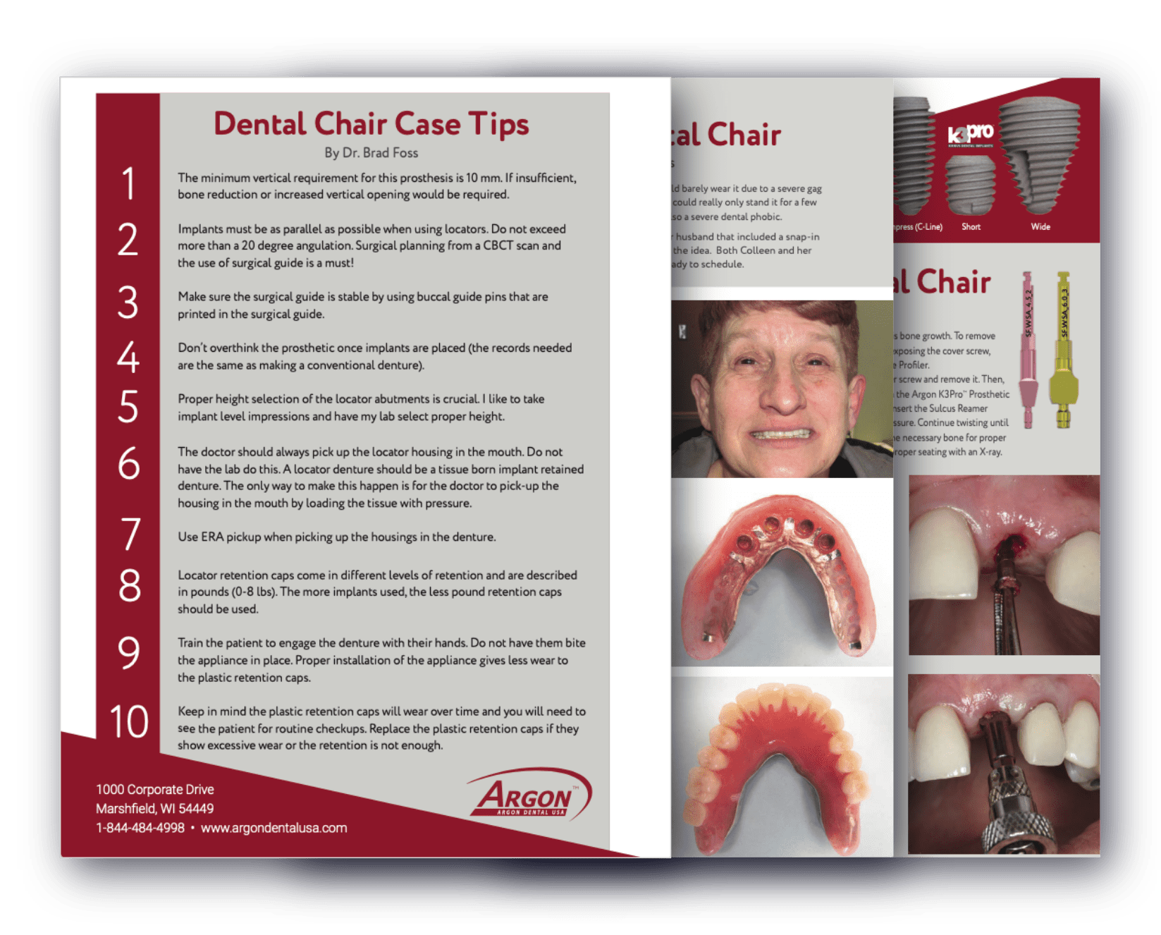
Case Studies
Case Spotlight: Minimally Invasive Implant Placement with Long-Term Stability
Clinician: Dr. Jan Bublik
Follow-Up Duration: 2 Years
System Used: Argon K3Pro™
Clinical Overview:
This case showcases a successful, minimally invasive approach to tooth extraction and single implant placement, with outcomes monitored over a two-year period.


Initial Presentation:
A preoperative radiograph revealed the clinical need for implant placement. Comprehensive planning was undertaken to ensure optimal esthetic and functional outcomes.
Atraumatic Extraction and Implant Placement:
The tooth was extracted with an atraumatic technique to preserve the surrounding bone and soft tissue. Immediately following extraction, a dental implant was placed, ensuring primary stability and ideal positioning.
Dual-Zone Grafting and Membrane Coverage:
To support both the soft and hard tissue contours, a dual-zone grafting technique was utilized. Surgical Esthetics collagen membrane was placed over the grafted socket to promote healing and protect the regenerative site.
Soft Tissue Management:
A simple suture technique was employed to ensure containment of the graft material and facilitate optimal soft tissue adaptation without excessive tension.
Follow-Up (2022):
At the 18-month post-operative mark, a follow-up radiograph demonstrated stable crestal bone levels and no signs of peri-implant bone loss, confirming the success of the biologically driven treatment approach.
Case Spotlight: Long-term success of a single implant restoration
Clinician: Dr. Meir
Follow-Up Duration: 4 Years
System Used: Argon K3Pro™
Clinical Overview:
This case illustrates the successful placement and long-term maintenance of a single dental implant with excellent esthetic and functional outcomes sustained over a four-year period.


Implant Surgery (2013):
The case began with the surgical placement of a dental implant after an immediate extraction, as seen in the intraoperative image.
Radiographic Confirmation (2013):
A post-operative periapical radiograph taken at the time of surgery confirmed correct implant positioning and osseous integration potential.
Early Healing Phase (2 Months Post-Placement):
Two months following placement, intraoral photographs revealed healthy, well-adapted soft tissue around the implant site, indicative of proper healing and early tissue integration.
Cosmetic Outcome (2014):
One year post-placement, the patient presented with excellent esthetic results. The final restoration harmonized seamlessly with the natural dentition.
Follow-Up X-ray One-Year Later (2015 ):
PA taken at two years demonstrated stable crestal bone levels with no evidence of bone loss.
Follow-Up X-ray Two-Years Later (2017):
Serial radiograph taken at four years post-operatively confirms a healthy implant, showing no evidence of bone loss.
Case Spotlight: Successful treatment of a failed root canal with a single implant
Clinician: Dr. Baldea
Follow-Up Duration: 3 Years
System Used: Argon K3Pro™
Clinical Overview:
This clinical case documents the placement and follow-up of a dental implant performed with radiographic and photographic records spanning three years, highlighting successful osseointegration and sustained peri-implant bone stability.


Pre-Operative X-Ray (2014)
The initial radiograph reveals a previously root-canaled tooth presenting with signs of periapical pathology. Evident radiolucency indicates an infection, suggesting failure of the endodontic treatment. Given the compromised state of the tooth and surrounding bone, extraction followed by implant placement was considered the most appropriate treatment plan. Despite the infection, there appeared to be sufficient bone volume to support future implant placement after adequate healing.
Pre-Operative Aesthetics:
A clinical image of the patient’s smile prior to implant placement was taken to document the aesthetic baseline and plan for optimal prosthetic integration, particularly regarding gingival contour and tooth color matching.
Surgical and Prosthetic Treatment:
In 2014, after healing of the extraction site, a dental implant was placed. The post-operative radiograph confirmed appropriate angulation and depth of placement with good primary stability.
One-Year Follow-Up (2015):
The radiograph showed stable peri-implant bone levels with no evidence of bone loss or peri-implant radiolucency, indicating successful osseointegration.
Two-Year Aesthetic Outcome (2016):
A photograph taken two years post-placement, revealed excellent soft tissue integration with harmonious gingival contours and color matching, contributing to a highly aesthetic result.
Three-Year Follow-Up (2017):
An x-ray taken three years post-placement showing no bone loss. Implant therapy can offer a predictable and long-lasting solution in cases of endodontic failure, especially when performed in a staged and carefully monitored approach. This case underscores the importance of comprehensive planning and follow-up in achieving both functional and aesthetic outcomes.